



Optical illusions reveal that vision is an active construction. The brain combines incomplete retinal signals with contrast, context, depth cues, motion, attention and prior experience to estimate what is in the world. An illusion exposes a situation in which those normally useful rules produce a stable perception that differs from a physical measurement.
Seeing is inference
The eye does not deliver a finished photograph. Photoreceptors sample light, retinal circuits compare nearby signals, the thalamus relays organized information, and multiple cortical areas respond to edges, color, motion, depth, objects and actions. Perception emerges through recurrent communication, not a single “vision center.” The result is fast and useful, but the image is constructed from evidence that can be ambiguous.
Consider brightness. The amount of light reaching a pixel-like retinal location depends both on an object’s reflectance and its illumination. A white page in shade may send less light to the eye than a dark surface in sunshine, yet we usually perceive stable materials. The visual system uses surrounding regions and assumptions about lighting. Designers of lightness illusions arrange context so those assumptions push identical patches toward different appearances.
Five families of useful illusion
| Family | What changes perceptually | What it probes |
|---|---|---|
| Brightness and color | Identical values appear different | Context, illumination and local contrast |
| Geometric | Equal lines or shapes appear unequal | Orientation, depth cues and spatial integration |
| Motion | A static image seems to move, or motion follows an unexpected path | Timing, eye movements and motion-sensitive circuits |
| Ambiguous figures | One image alternates between interpretations | Competition, attention and figure–ground assignment |
| Illusory contours | An edge or surface appears where none is drawn | Filling-in and boundary completion |
Edges that are not on the page
In a Kanizsa-style figure, carefully arranged “pac-man” shapes make viewers perceive a bright triangle with crisp borders, despite no triangle outline being present. Brain-imaging and electrophysiology studies indicate that early visual areas, including V2 and in some contexts V1, participate in representing subjective contours. Higher areas contribute feedback. The lesson is not that one patch of cortex invents the triangle; boundary completion is distributed across levels.
Why identical colors look different
Simultaneous contrast makes a gray patch on a dark field appear lighter than the same gray on a light field. More elaborate displays add inferred shadow or illumination. Viral demonstrations often invite people to argue about the “true” color. Physical measurements can establish the light reaching the display, while perceptual reports describe how the visual system interprets it. Both questions are legitimate, but they are not interchangeable.
Motion from stillness
Some static patterns appear to ripple during small eye movements. Unequal processing delays for different brightness transitions can contribute. Apparent motion offers an even simpler principle: show stimuli at separate positions in the right sequence and the observer experiences movement between them. Functional imaging has found activity along an illusory motion path in early visual cortex, showing that neural activity can track perceived rather than physically continuous motion.
Two answers in one image
The Rubin vase can be seen as a central vase or as two facing profiles. The retinal stimulus stays fixed while figure and background swap. Necker cubes reverse depth orientation. Such bistable images show that perception settles on one coherent interpretation at a time, then may switch as adaptation, attention and competition change. They do not reveal whether a viewer is “left-brained,” creative or troubled; personality claims attached to illusions are usually unsupported.
- Light is sampled by rods and cones.
- Retinal networks emphasize contrast and change.
- Signals are organized into maps and features.
- Context and prior experience constrain possible scenes.
- Competing interpretations settle into a useful percept.
- New evidence or attention can trigger a different interpretation.
Illusions are features of a capable system
Calling every illusion an error misses the larger achievement. The visual system must infer three-dimensional, stable objects from shifting two-dimensional images, blind spots, eye movements and changing light. Rules that succeed across natural environments can be exposed by artificial arrangements. An illusion is often the price of using efficient assumptions rather than recomputing reality from nothing.
What varies between people
Illusion strength can vary with age, visual experience, attention, culture, eye conditions and neurological differences. Screens add uncontrolled variables: brightness, color calibration, viewing distance and image compression. A single online illusion is not a diagnostic test. Persistent new visual symptoms deserve professional assessment; a fun image cannot confirm or exclude a medical condition.
- Vision is a layered inference, not a direct copy of the world.
- Context helps perception remain stable but can produce predictable mismatches.
- Illusions let researchers separate the physical stimulus from subjective appearance.
- Early and higher visual areas interact; there is no single illusion switch.
- Online illusions should not be used to diagnose personality or health.
- Ffytche & Zeki, brain activity related to illusory contours.
- Muckli et al., PLoS Biology, primary visual cortex and apparent motion.
- Weidner & Fink, neural correlates of geometric illusions.
- NIH/PubMed Central reviews on visual illusions and constructive perception.
Why a retinal image cannot specify the world
Every retinal image is compatible with many possible scenes. A small object nearby and a large object far away can cast the same outline. A gray surface under bright light and a white surface in shadow can send similar light to the eye. Motion on the retina might come from an object, the observer’s eyes or the observer’s body. Vision succeeds by combining incoming signals with regularities learned from the physical world.
This is sometimes described as unconscious inference or predictive processing. The terms belong to several theories that differ in detail, so an illusion should not be presented as proof of one grand model. The secure point is narrower: perception uses context and internal processing to resolve ambiguity. Carefully designed images let researchers hold the physical stimulus constant while changing what observers report.
An illusion is also not the same as a hallucination. An illusion begins with an external stimulus that is interpreted in a systematically surprising way. Hallucination refers to a perceptual experience without the corresponding external stimulus. The categories can become clinically complex, but a normal geometric illusion does not indicate that a viewer is hallucinating.
The retina edits before the brain sees
Rods and cones convert light into neural signals. Horizontal, bipolar and amacrine cells combine information, and retinal ganglion cells send output through the optic nerve. Center-surround receptive fields emphasize differences between neighboring regions rather than reporting absolute brightness independently. This helps detect edges across changing illumination and contributes to simultaneous contrast effects.
The optic nerve creates a blind spot where ganglion-cell axons leave the eye, yet people do not normally see a hole. The visual system uses information from the other eye and surrounding patterns to fill a coherent percept. A simple blind-spot demonstration reveals the process, but “fill in” does not mean the brain paints a tiny internal screen. It means neural activity supports a continuous experience despite missing retinal samples.
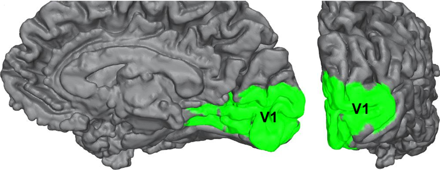
Signals pass through the lateral geniculate nucleus and reach primary visual cortex, or V1, in the occipital lobe. Neighboring points in visual space map to neighboring cortical regions. Neurons respond selectively to orientation, spatial frequency and other features. Processing then continues through interconnected cortical streams involved in object identity, space and action. Feedback runs toward earlier stages as well as forward.
Brightness is a judgment about illumination
In simultaneous contrast, identical central squares appear lighter or darker depending on their surrounds. Lateral interactions contribute, but more complex lightness illusions cannot be explained by local contrast alone. The visual system estimates surfaces, shadows and lighting. A checker-shadow display is powerful because cues suggest that one patch sits in shade, prompting perceptual compensation.
Digital demonstrations should include a measurement check. A color-picker can confirm that two image regions have equal pixel values, while a bridge placed between them reduces contextual separation. This does not prove one viewer’s experience is wrong. It demonstrates that physical equality at the screen and perceived lightness answer different questions.
Screen settings matter. Automatic brightness, high-dynamic-range processing, viewing angle and compression can alter pixels. Barnakle diagrams should be original, labelled as demonstrations, and accompanied by instructions that do not rely on perfect color reproduction.
Color constancy and famous disagreements
Color perception depends on the spectrum of light, cone responses and the brain’s estimate of illumination. Color constancy helps a familiar object appear relatively stable indoors, outdoors or in shade. Ambiguous photographs can leave the illuminant uncertain, leading different viewers to discount different colors and report strikingly different appearances.
The lesson is not that color is imaginary. Wavelength distributions and display values are measurable, and color matches can be tested systematically. Perceived color is a biological response to those signals in context. Language, adaptation and individual differences in lenses and photopigments also influence reports.
Afterimages reveal adaptation. Stare at a strongly colored pattern, then shift to a neutral field, and opponent channels respond out of balance. The temporary complementary appearance shows that current perception depends partly on recent stimulation. It is normally brief; persistent or unexplained visual changes are a medical question, not an online personality test.
Contours the image never drew
Kanizsa figures arrange incomplete disks and angles so observers perceive an occluding surface with boundaries. The visual system favors an interpretation in which one coherent shape blocks several objects. Neuroimaging studies associate illusory contours with activity in visual cortex, including V2 and context-dependent responses in V1.1
Researchers compare an illusory-contour display with control arrangements containing similar local elements but no coherent shape. If activity differs with reported contour, the result is harder to attribute to the individual pieces alone. Timing methods can ask when early and later regions participate, while brain stimulation and lesion evidence can test causal roles.
Boundary completion is useful outside the laboratory. Objects are frequently hidden behind branches, furniture or other people, yet perception treats visible fragments as one continuing object. The illusion isolates a rule that usually prevents the world from appearing shattered.
Apparent motion and the cinema principle
Two lights flashed at nearby locations in sequence can produce a percept of one light moving. Film and animation exploit related temporal integration, although real cinema perception involves many cues beyond the simplest phi phenomenon. The brain uses timing and position to infer a continuous event from discontinuous samples.
Functional imaging has found activity in V1 along a path where motion was perceived even though no stimulus occupied the intervening locations.2 Feedback from motion-sensitive higher regions may help sustain that representation. This is an important example of why a strictly feed-forward ladder is incomplete.
Static motion illusions use asymmetric luminance, repeated patterns and small eye movements. Different designs rely on different mechanisms. Saying “your peripheral vision is broken” is not an explanation; researchers manipulate contrast, adaptation, fixation and timing to identify which variables drive the effect.
Size and length depend on context
In the Müller-Lyer illusion, equal line segments appear unequal when arrow-like fins point inward or outward. In the Ebbinghaus illusion, a central circle appears larger or smaller depending on surrounding circles. These displays show that spatial judgments integrate neighboring geometry. The strength varies across viewers and versions.
One explanation links some geometric illusions to depth and perspective cues learned in built environments. That cannot account for every result by itself. Low-level orientation interactions, spatial filtering, attention and decisional factors also contribute. Modern research treats the illusion as an outcome with multiple levels rather than a single trick.
Action and perception can respond differently in some tasks. A person’s conscious size estimate may show an illusion more strongly than a rapid grasping movement, though experimental findings depend on feedback and design. This supports partially specialized visual pathways without dividing the brain into a simple “seeing system” and an immune “doing system.”
Ambiguous figures reveal competition
The Rubin vase alternates between a vase and two profiles because one region is assigned as figure while the other becomes background. A Necker cube flips between depth orientations. The stimulus does not change, and viewers usually cannot hold both interpretations with equal stability. Neural populations representing alternatives compete while attention and adaptation bias the outcome.
Bistable perception gives researchers repeated perceptual changes without changing the image. Participants report switches while brain activity is recorded, allowing investigators to look for signals associated with subjective interpretation rather than stimulus onset. Reporting itself adds motor and decision activity, so no single activated area can be labelled “the place perception happens.”
Rates of switching vary among people and with attention, but they are not a reliable personality diagnostic. Online posts claiming the first image seen reveals creativity, trauma or a brain hemisphere typically lack validated evidence.
Faces: a system tuned to socially important patterns
Pareidolia is the experience of seeing a meaningful pattern, often a face, in clouds, outlets or rocks. Faces are important and share a common arrangement, so a sensitive detector that occasionally produces false positives may be useful. Brain studies show face-like objects can recruit portions of the face-processing network, while observers still know they are looking at an object.
This differs from a delusional belief that an object is literally a person. Ordinary pareidolia is widespread. The scientific opportunity is to vary how face-like a pattern is and measure detection, attention and neural response.
Illusions across development and culture
Children’s susceptibility changes as visual systems, attention and experience develop. Aging alters optics and neural processing. Cataracts, retinal disease and neurological conditions can change perception, but group differences do not turn a recreational image into a diagnostic instrument. Clinical tests require standardization, comparison populations and evidence about sensitivity and specificity.
Cross-cultural studies have reported variation in some geometric illusions, possibly related to visual environments and learned interpretation. Sampling and measurement matter: differences within cultures can exceed group averages, and exposure to images or built environments changes. The responsible conclusion is that visual rules combine shared biology with experience, not that cultures possess fundamentally different realities.
How scientists use illusions as controlled disagreements
Psychophysics measures the relation between physical stimuli and reported perception. Researchers vary one property, randomize trials, include controls and fit thresholds or bias estimates. Eye tracking establishes fixation. EEG and MEG supply timing; MRI supplies spatial patterns; recordings in animal models examine cells and circuits. Causal tools can perturb activity. Converging methods matter because each has limits.
A beautiful demonstration may be large but scientifically ambiguous. A less dramatic stimulus can isolate a variable and support stronger inference. Researchers preregister analyses, share code and replicate effects to distinguish robust mechanisms from flexible storytelling.
An original Barnakle contrast test
Build: place two identical mid-gray circles on separate rectangles, one near-black and one near-white. Add a button or printed strip that visually connects the circles.
- Compare the circles while separated by their surrounds.
- Reveal the connecting strip with the same gray value.
- Measure the digital values with a color picker.
- Record whether knowing the values removes the appearance.
Most viewers continue to experience some difference even after learning the circles match. Knowledge and perception can disagree without either being irrational. The visual system’s contextual operations are automatic, while measurement answers a deliberate physical question.
How to evaluate an illusion claim
- Verify the image itself has not changed through animation, compression or adaptive display settings.
- Identify the measurable physical equality or mismatch.
- Look for a controlled comparison rather than an anecdote.
- Separate a proposed mechanism from an established observation.
- Reject personality or medical claims unless supported by validated research.
Illusions deserve their popularity. They let a person experience a research question directly. Their deepest lesson is not that vision is untrustworthy, but that accurate everyday seeing requires interpretation. The world reaches the brain as evidence; perception is the remarkably effective model built from it.
Numbered references
- Ffytche D.H. & Zeki S. “Brain activity related to the perception of illusory contours.” NeuroImage (1996). PubMed.
- Muckli L. et al. “Primary visual cortex activity along the apparent-motion trace reflects illusory perception.” PLoS Biology (2005). doi:10.1371/journal.pbio.0030265.
- Weigelt S. et al. “Seeing things: illusory contours in the human visual brain.” Journal of Neuroscience. PMC.
- Grossberg S. “How visual illusions illuminate complementary brain processes.” Trends in Cognitive Sciences (2014). PMC.
Keep exploring.
One remarkable idea at a time—nature, science, history and beyond.
Sources and further reading
Barnakle uses credible primary and authoritative sources wherever possible.
- Ffytche & Zeki 1996 illusory contoursnMuckli et al. PLoS Biology 2005 apparent motionnNIH/PubMed Central illusion reviews
Last reviewed September 12, 2026.

Barnakle Editorial Team
A member of the Barnakle editorial team, exploring remarkable ideas with clarity, curiosity and care.
More from this author →